
Odds are you’ve heard of an MRI, but may not know how it works unless you are a doctor. A MRI, which stands for Magnetic Resonance Imaging, is a commonly used diagnostic test in medicine. Although it is commonly used, many people do not know how it works or why they need one. This is a beginner’s guide to understanding an MRI.
What exactly is an MRI?
First and foremost, what does MRI even stand for? The answer is Magnetic Resonance Imaging. An MRI is a type of scan that doctors use to view an area such as a person’s head, chest, leg or whatever is needed. As is in the name, through the use of magnets in combination with radio waves, images of the inside of the body are created. This allows doctors to view organs, tissues, and even skeletal system all without needing to make any cuts to look inside.
How does an MRI work?
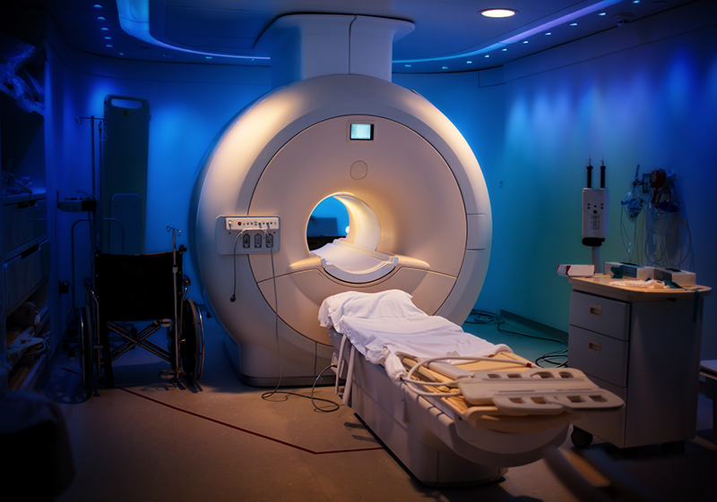
The way an MRI works is that you lie down on a moveable table, and it slides you into this tube that is open at both ends, and is surrounded by magnets. The reason this works to produce images is because humans are made primarily of water. A molecule of water consists of 2 hydrogen atoms and an oxygen atom. During an MRI, the radio waves cause the hydrogen atoms to realign for a short time. When the atoms begin falling back into their natural places, they give off a signal. The magnets in the machine are able to pick up these faint signals, and then a computer is able to convert the signals into images that your doctor can read.
What do doctors use an MRI for?
Doctors use MRI’s for diagnostic purposes to be able to see and recognize medical issues or healing processes in patients. An MRI is non-invasive which means the doctors do not have to actually make an incision to see what they are dealing with. Doctors can use an MRI for imaging for a lot of the human body.
Why may you need an MRI and what can it show?
You may need an MRI because it can help doctors diagnose the issue or see how progress is coming along with treatment to make sure everything is healing as it should. The primary uses of an MRI are checking the brain, spine, heart, blood vessels, bones, and joints. MRIs can also be used to check a woman’s breasts and ovaries, a man’s prostate, along with a person’s liver, kidneys and pancreas.
An MRI on your brain and spinal cord can show evidence of blood vessel damage, cancer, multiple sclerosis, stroke, and brain and/or spinal cord injuries. MRI of the heart and blood vessels can show damaged caused by a heart attack, heart disease, blocked blood vessels and even problems dealing with the heart’s structure. You can also get an MRI on your bones and joints. This can show problems with cancer, bone infections, spinal disc problems, and overall damage to joints.
How does an MRI differ from a CT scan?
Both an MRI and CT scan provides diagnostic images, but there are differences. For one, a CT scan uses x-rays whereas an MRI uses magnets and radio waves. Another difference is that a CT scan gives of a tiny amount of radiation, but an MRI does not. The x-rays for a CT scan need a tiny bit of ionizing radiation in order to get the images which is why radiation is given off. A CT scan can also be done much quicker than an MRI can, as they may only take 5 minutes.
What do you do during an MRI and how long should it take?
The time it takes to get an MRI varies due to how many images are needed, and what part of the body is being examined. That said, the process of getting an MRI usually takes between 20 minutes and 1 hour. The MRI images are also taken in sequences that last only a few seconds or minutes. This means you aren’t forced to be still for the entire length of the exam, but it is best if you do not make drastic movements. During the imaging sequences, it is important to be as still as possible while the MRI is going on. The more movement there is the more likely it is that the image is not clear and it has to be done again.
Is there a difference between an open and closed MRI?
Most MRI machines are “closed” as it has a top and bottom so it is like being inside of a tube. Being stuck in a small tube can understandably be claustrophobic. To solve this, some places offer what is called an “open” MRI. What this means is that instead of completely surround the person, the magnets are just on the top and bottom, allowing the sides to be open.
During an MRI why can’t you have any metal on you?
This is a pretty easy question. Simply, the magnets in the machine are powerful, so if you have any metal on you it will get sucked to the magnet. This can cause damage to the machine and to the person inside of it. This is why a lot of times doctors will ask you to change into a hospital gown before you get an MRI.
What should you disclose to your doctor before your MRI?
Since an MRI is using powerful magnets, there are some risks. If you have a metallic joint or joints, artificial heart valve,implantable heart defibrillator,pacemaker, aneurysm clips, cochlear implants, tattoos, piercings, a bullet, shrapnel, or any other sort of metal, let your doctor know so they can make a decision as to whether it will be affected by the MRI.
Sources:
https://www.mayoclinic.org/tests-procedures/mri/about/pac-20384768
https://www.webmd.com/a-to-z-guides/what-is-an-mri#1
https://www.radiologyinfo.org/en/info.cfm?pg=bodymr
https://www.medicalnewstoday.com/articles/146309.php
https://osmri.com/open-vs-closed-mri/
https://www.viaradiology.com/news/whats-difference-ct-scan-mri/


0 comments
Write a comment